Machine Learning for Infection and Disease (MLID)
The MLID Group (Yakimovich Lab) is a Young Investigator research group at the Center for Advanced Systems Understanding (CASUS) / Helmholtz-Zentrum Dresden-Rossendorf (HZDR) in Görlitz, Germany.
We work at the intersection of Machine Learning, Computational Microscopy, and Infection Biology, pioneering algorithmic methods to understand, quantify, and model how pathogens interact with host cells.
What We Are Working On
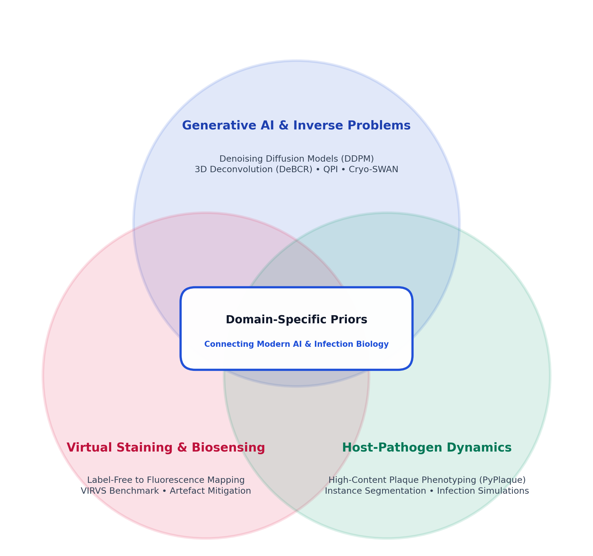
We connect these research domains through the incorporation of domain-specific physical and biological priors into modern AI techniques. By grounding data-driven models in the physical principles of optical wave propagation, image formation mechanics, and host-pathogen molecular dynamics, our approaches achieve high biological fidelity, generalisation, and physical interpretability.
1. Generative AI & Physics-Informed Inverse Problems
Standard light microscopy produces noisy, diffraction-limited, and degraded measurements. We design generative models (including denoising diffusion probabilistic models, conditional variational diffusion, and Beylkin-Coifman-Rokhlin neural networks) constrained by physical imaging models to solve fundamental inverse problems:
- Deep Microscopy Deconvolution: Reconstructing 3D in-focus widefield and super-resolution microscopy volumes.
- Single-Exposure Quantitative Phase Imaging (QPI): Inferring quantitative phase representations from conventional microscopes using generative priors.
- Cryo-EM & Molecular Volume Density Representations: High-resolution representations of macromolecular complexes (Cryo-SWAN).
2. Virtual Staining & Computational Biosensing
Fluorescence staining and genetic reporter tagging allow specific pathogen detection but often cause phototoxicity, alter cellular physiology, and require invasive preparation. We develop Virtual Staining methodologies:
- VIRVS (Virus Infection Reporter Virtual Staining): Mapping continuous label-free brightfield micrographs directly to multi-channel fluorescence infection reporter signals without physical fluorophores.
- Deep Learning Quality & Artefact Detection: Autonomous quality control, focus classification, and artefact mitigation in high-content screening.
3. Phenotypic Profiling & Host-Pathogen Dynamics
Viruses remodel host cell morphology and organelle organization throughout their life cycle. We develop open-source automated computer vision systems and quantitative phenotyping pipelines:
- High-Throughput Plaque & Colony Phenotyping: Tools such as PyPlaque for high-throughput, unbiased quantification of viral fitness and infectivity.
- Biomedical Instance Segmentation: Star-convex polygon models and weak-supervision frameworks tailored for dense, spatially-correlated infectious microenvironments.
- Cellular Dynamics & Infection Simulation: Bridging cellular microscopy observations with tissue-scale computational simulations.